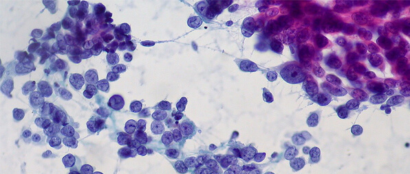
Fluids, Urine, and Effusions Cytology
Our firm proudly offers specialized cytology and histopathology services, both crucial for diagnosing diseases at the cellular level and guiding effective treatment.
Our skilled team of pathologists provides unparalleled cytology services, encompassing the meticulous analysis of fluids, urine, and effusions. Employing cutting-edge techniques, we conduct comprehensive urine, pleural fluid, ascetic fluid, and synovial fluid tests. Through precise cellular morphology assessment and advanced staining methodologies, we identify abnormal cells and differentiate cellular types to ensure accurate diagnoses, effectively managing various clinical conditions.
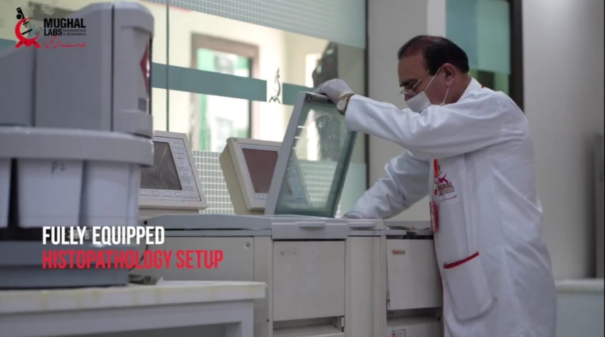
Simultaneously, we specialize in histopathology, ensuring the accurate analysis of tissue samples. A strong understanding of biology is applied to interpret complex data, as histopathology reports are essential for determining the stage and severity of diseases like cancer.
With a commitment to excellence and a focus on advancing medical diagnostics, our laboratory ensures you receive top-tier fluid cytology and tissue histopathology services for optimal patient care. Let’s connect for better testing.