https://www.youtube.com/watch?v=8O8ltfOuP6s

Immunohistochemistry (IHC) Studies
Immunohistochemistry (IHC) is currently used in both research and clinical settings for the diagnosis of abnormal cells and the distribution and localization of certain proteins of interest in the cell. We offer Lab Testing and Diagnostics services including IHC staining services using a wide range of validated antibodies– over 130 antibodies for a variety of pathological markers. Furthermore, listed below are commonly used and available IHC panels:
Read More
BREAST (Beta-Catenin, Calponin, Cytokeratin 14, Cytokeratin AE1/AE3, E-Cadherin, EMA, Estrogen Receptor, GATA3, GCDFP-15, HER-2 neu, Ki-67, Mammaglobin, Myosin Smooth Muscle Heavy Chain, p120, p53, p63, Progesterone Receptor, SMA)
CARCINOMA OF UNKNOWN PRIMARY SITE (CD10, CDX2, CK19, CK5/6, Cytokeratin 20, Cytokeratin 7, GATA3, GCDFP-15, HepPar1, Napsin , PAX8, PSA, Thyroglobulin, TTF-1, WT1)
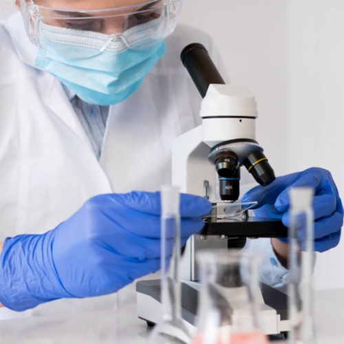
Mughal Labs is renowned for its advanced lab testing and diagnostic services, ensuring precise and reliable results. Also, our facility provides a comprehensive range of advanced lab testing and diagnostic services to meet diverse medical needs. Mughal Labs offers comprehensive lab testing and diagnostic services to ensure accurate and timely results.
Our clinic specializes in high-quality lab testing and diagnostic services for a wide range of medical conditions. We provide reliable lab testing and diagnostic services, utilizing the latest technology and expert analysis.
Flowcytometry
Flow cytometry is a method for analyzing the expression of cell surface and intracellular molecules, characterizing and defining different cell types in a heterogeneous cell population. Also, it allows simultaneous multi-parameter analysis of single cells. It is a powerful tool for interrogating the phenotype and characteristics of biological cells.
Moreover, it is based upon the light-scattering properties of the cells being analyzed and these include fluorescence emissions. This fluorescence may be associated with dyes or conjugated to monoclonal antibodies specific for molecules either on the surface or in the intracellular components of the cell. Likewise, Flow cytometry facilitates the identification of different cell types.
All forms of cytometry depend on the basic laws of physics, including those of fluidics, optics, and electronics. Flow cytometry is a system for sensing cells or particles as they move in a liquid stream through a laser (light amplification by stimulated emission of radiation)/light beam past a sensing area. Moreover, the relative light-scattering and color-discriminated fluorescence of the microscopic particles is measured and displayed using histograms.
Flow cytometers are multi-parameter, recording several measurements on each cell; therefore, it is possible to identify a homogeneous subpopulation within a heterogeneous population. Likewise, this is one of the most useful features of flow cytometers and makes them preferable to other instruments such as spectrofluorimeter, in which measurements are based on analysis of the entire population.
In clinical laboratories, this technology is used to quickly and accurately diagnose various types of leukemia and lymphomas. With a focus on innovation, Mughal Labs delivers top-tier advanced lab testing and diagnostic services for accurate diagnoses. We are committed to excellence in advanced lab testing and diagnostic services, utilizing the latest technology and expertise.


Research & Development
Research and development is an important element of our core motive. We constantly strive to facilitate new research projects of both clinical and academic nature in order to put our contribution into scientific advancements, ultimately leading to better diagnosis of disease and patient management.
Our team comprises highly qualified medical personnel with extensive experience in research and development with a large list of published research studies in both local and international journals associated with their names.
Our team comprises highly qualified medical personnel with extensive experience in research and development with a large list of published research studies in both local and international journals associated with their names.
At the moment, efforts are being put to develop a full-fledged Clinical research office with a pool of trained research staff, scientists, and statisticians to ensure a unique combination of expertise.
Currently, we’ve collaborated and offered our services for various different projects in the field of Histopathology including grossing, processing, embedding, and staining and ancillary tools such as special stains and an extensive panel of Immunohistochemical stains. Our collaborators include local, national and international partners. Experience exceptional care with our advanced lab testing and diagnostic services, designed to support effective medical evaluations.
With our advanced lab testing and diagnostic services, you can trust us for precise and thorough evaluations. At Mughal Labs, our lab testing and diagnostic services are designed to support effective patient care and treatment planning.
Polymerase Chain Reaction (PCR)
The polymerase chain reaction (PCR) is a nucleic acid based testing to provide diagnostic information to clinicians in order to help guide optimal care for patients with cancer, viral infection, histocompatibility and genetic diseases.
It is a scientific technique in molecular biology to amplify a single or a few copies of a piece of DNA across several orders of magnitude, generating thousands to millions of copies of a particular DNA sequence. The polymerase chain reaction is a powerful technique that has rapidly become one of the most widely used techniques in molecular biology because it is quick, inexpensive and simple. The technique amplifies specific DNA fragments from minute quantities of source DNA material.
From the daily practicalities of medical diagnosis to the theoretical framework of systematics, from courts of law to field studies of animal behavior, PCR takes analysis of tiny amounts of genetic material-even damaged genetic material to a new level of precision and reliability.
Currently, we are providing PCR testing for the following:
- COVID-19 PCR (Including Delta variant)
- HCV PCR (Qualitative and Quantitative)
- HBV PCR (Qualitative and Quantitative)
- Dengue RNA by PCR
- HIV by PCR (Viral Load/Quantitation)
- MTB by PCR
- HLA B27 by PCR


Special Staining Services
Special stains traditionally refer to any staining other than an H&E. It covers a wide variety of methods that may be used to visualize particular tissue structures, elements, or even micro-organisms not identified by H&E staining. Listed below are several special stains performed on regular basis:
- Giemsa (micro-organisms)
- Gomori’s Methenamine Silver or GMS (fungi, basement membrane)
- Masson’s Trichrome (muscle, fibrin, connective tissue)
- Periodic Acid Schiff’s or PAS /PASD (fungi, carbohydrate, glycogen)
- Perl’s Prussian Blue (hemosiderin, iron)
- Ziehl-Neelsen or ZN (acid fast bacilli, TB)
- Congo Red stain (amyloid)
- Mucicarmine stain (mucin)
- Oil red O stain (fat)
- Reticulin stain (reticulin meshwork)